New Dry Eye Technology at Bridge Eye Care
At Bridge Eye Care, we’re always looking for ways to better understand what is happening with your eyes, not just whether you can see clearly, but how healthy and comfortable your eyes are.
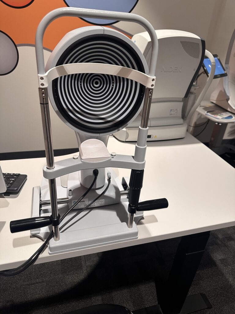
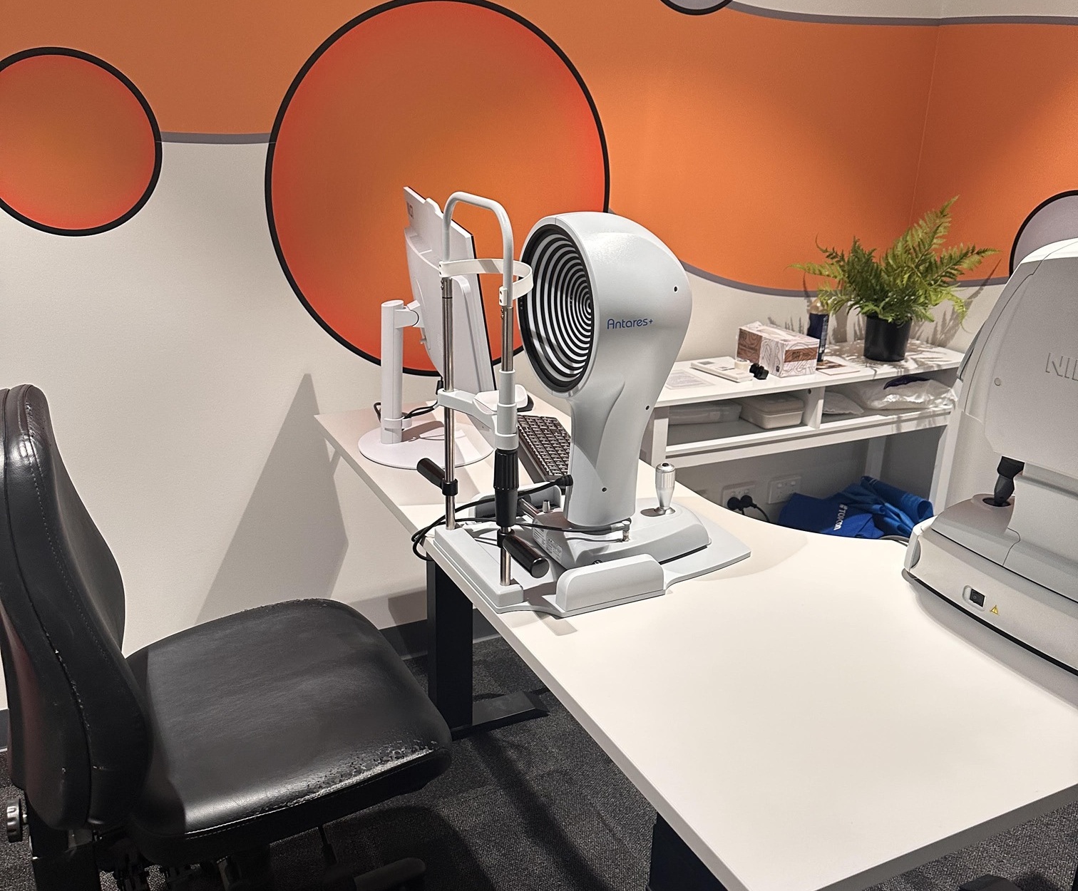
We’re excited to introduce our new ANTERES corneal topographer and meibographer, advanced technology that gives us an even more detailed view of the front surface of the eye and the tiny oil-producing glands within the eyelids.
Mapping the surface of your eye
The cornea is the clear front surface of the eye. The ANTERES creates a detailed map of its shape and curvature, helping us identify subtle irregularities that may not be visible during a standard eye examination.
This can be useful when assessing corneal conditions, fitting contact lenses and investigating changes to the eye’s surface associated with dry eye.
The imaging is quick, comfortable and completely non-invasive.
A Closer look at dry eye
One of the features we’re particularly excited about is meibography. Dry eye is much more complex than simply “not producing enough tears”.
Inside our upper and lower eyelids are tiny structures called meibomian glands. These produce the oily component of our tears, which helps prevent tears from evaporating too quickly.
When these glands become blocked or don’t function properly; known as meibomian gland dysfunction (MGD), it can contribute to dry, gritty, burning or watery eyes, redness, fluctuating vision and difficulty wearing contact lenses.
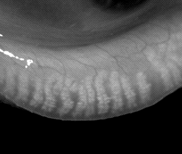
We can now actually see your meibomian glands
Using infrared imaging, the ANTERES allows us to take detailed images of the meibomian glands hidden inside your eyelids. We can assess their structure and look for changes such as shortening, distortion or gland loss.
Importantly, we can show you the images of your own glands. This can make it much easier to understand what may be contributing to your symptoms and why we have recommended a particular treatment.
Why does this matter?
There isn’t one single type of dry eye, and there isn’t one treatment that is right for everybody. Two people can have very similar symptoms but quite different underlying causes.
By combining your symptoms with a careful eye examination and advanced imaging, we can better identify the factors contributing to your dry eye and develop a treatment plan specifically for you. It also gives us objective information that we can document and refer back to when monitoring your eye health over time.
Building our dry eye service at Bridge Eye Care
The addition of the ANTERES is part of our ongoing development of a more comprehensive approach to dry eye assessment and treatment in Murray Bridge. Our aim is to move beyond simply recommending eye drops for uncomfortable eyes. Instead, we want to understand why your eyes are dry, identify the underlying factors where possible, and then recommend treatment accordingly.
And, as with all the technology we use at Bridge Eye Care, the equipment is only one part of the picture. The most important part is interpreting the information in the context of you, your symptoms and your eyes.
If you experience dry, watery, gritty, red or uncomfortable eyes; or vision that seems to fluctuate throughout the day, talk to our optometrists about a dry eye assessment at Bridge Eye Care in Murray Bridge.